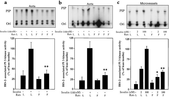
Figure 4.
IRS-1– and IRS-2–associated PI 3-kinase activities in the aorta and microvessels ex vivo. Isolated aortas were incubated without or with insulin (100 nM) in DMEM (0.1% BSA) for 30 minutes at 37°C. Equal amounts of protein (2 mg) samples were subjected to immunoprecipitation with αIRS-1 (a) or αIRS-2 (b) antibodies. In the studies with microvessels (c), samples were treated without or with insulin (2–100 nM) in DMEM (0.1% BSA) for 5 minutes at 37°C, and 2-mg protein samples were subjected to immunoprecipitation with αIRS-2 antibody. The kinase activities were measured in the presence of phosphatidylinositol and [32P]ATP, and the lipid products were separated by TLC (top panels). The spots that comigrated with a PI 3-phosphate (PIP) standard were quantified by a PhosphorImager. Data (mean ± SD; n = 4) are expressed as relative to control, assigning a value of 100% to the mean of 100-nM insulin-treated samples of lean rats. *P < 0.05, **P < 0.01, lean vs. obese treated with insulin at the same concentrations.

