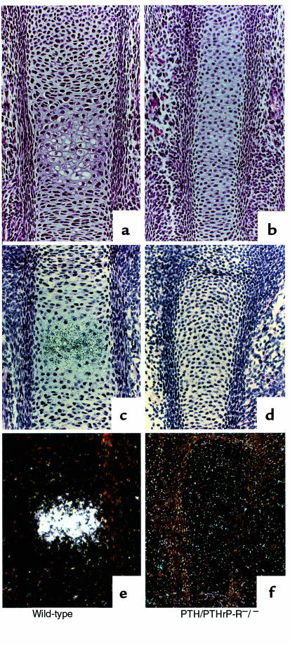
Figure 1.
Chondrocyte differentiation at E15.5. Hematoxylin/eosin staining (a and b) and type X collagen mRNA in situ hybridization (c–f) of wild-type and PTH/PTHrP-R–/– phalanges. PTH/PTHrP receptor–ablated bones show a delay in chondrocyte differentiation and type X collagen expression (b, d, and f) when compared with wild-type bones (a, c, and e). Bright-field, c and d; dark-field, e and f.

