
Figure 3.
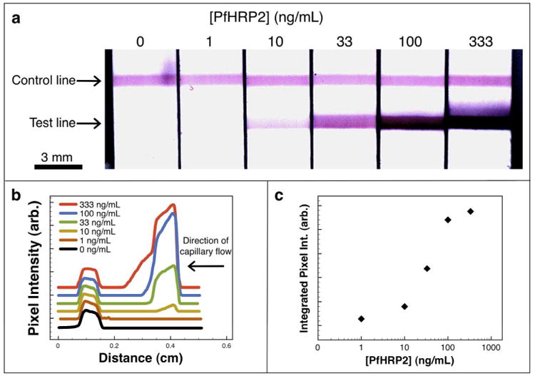
Recombinant PfHRP2 immunoassay. (a) Flow strip images, (b) green channel pixel intensity line scans offset along the y-axis for clarity, and (c) the corresponding sigmoidal curve generated by a typical magnetic enrichment immunoassay performed on recombinant PfHRP2 biomarkers in spiked human plasma. The starting sample of 500 μL was magnetically enriched 50-fold prior to analyte visualization by immunochromatography.