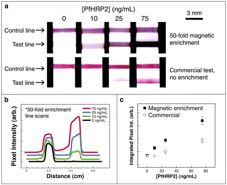
Figure 4.

Comparison of magnetic enrichment and commercial assay. (a) Flow strip images from a 50-fold magnetic enrichment immunoassay (top row), and from the unmodified commercial assay performed with no enrichment (bottom row). (b) Green channel pixel intensity line scans for the magnetically enriched samples offset along the y-axis for clarity. (c) The integrated green channel pixel intensity at the test line plotted as mean ± standard deviation (n=3) for the two assay systems.
