Figure 1.

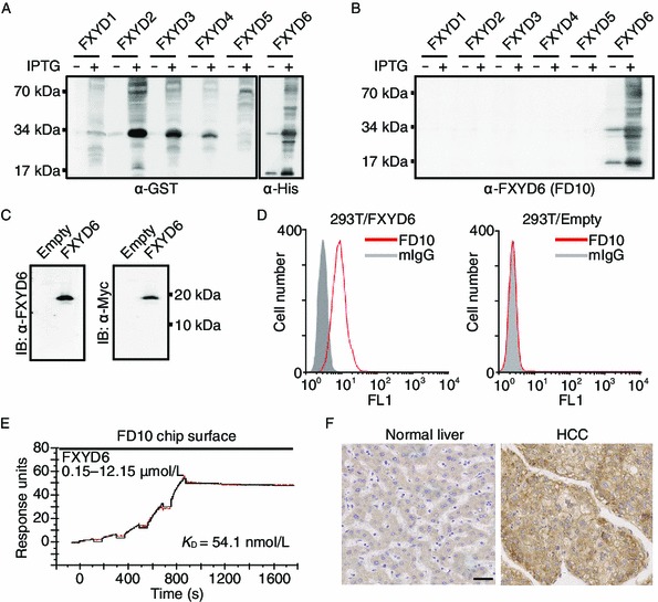
Anti-FXYD6 mAb of FD10 possesses high specificity and affinity. (A and B) Bacteria-expressed FXYD1–6 proteins from whole cell lysate without or with IPTG were detected by anti-Tag (A) or anti-FXYD6 mAb of FD10 (B). (C) Immunoblotting analysis of FXYD6 expression in 293T cells. Cells were transfected with pcDNA3.1-empty or pcDNA3.1-FXYD6, and whole cell lysate was analyzed using anti-FXYD6 antibody of FD10 (left panel) or anti-myc antibody (right panel). (D) FACS analysis of FD10 binding with FXYD6. The 293T cells, transfected with pcDNA3.1-FXYD6 (left panel) or with pcDNA3.1-empty (right panel), were incubated with FD10 or control normal IgG. (E) SPR sensorgrams illustrating binding of FD10 to FXYD6 proteins (black line) and fitted saturation binding curves (red line). The dissociation constant (KD) was calculated from the saturation binding curve. (F) Normal liver tissue (left panel) and HCC tissue (right panel) were stained by immunohistochemistry with FD10 (bar, 40 μm)
