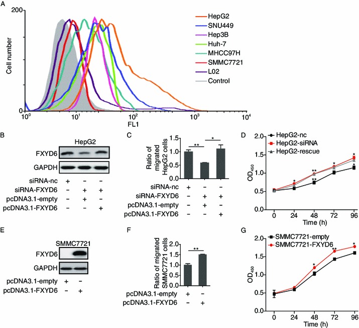
Figure 2.

FXYD6 promotes the migration and proliferation of HCC cells. (A) FACS analyzed the expression of FXYD6 protein within 6 HCC cell lines and L02 control cell line using FD10. Control cells were SMMC7721 stained by mIgG. (B) Immunoblotting of FXYD6 protein in HepG2 cells after 36 h co-transfection with siRNA-FXYD6 and pcDNA3.1-FXYD6. (C) The migration of HepG2 cells was determined after 36 h co-transfection with siRNA-FXYD6 and pcDNA3.1-FXYD6. *P < 0.05 or **P < 0.01, compared with the siRNA-FXYD6 group. (D) The proliferation of HepG2 cells was determined after co-transfection with siRNA-FXYD6 and pcDNA3.1-FXYD6 at different time points. Rescue assay was conducted in HepG2 cells co-transfected with siRNA-FXYD6 and pcDNA3.1-FXYD6. *P < 0.05 or **P < 0.01, compared with the siRNA-FXYD6 group. (E) Immunoblotting of FXYD6 protein in SMMC7721 cells after 24 h transfection with pcDNA3.1-FXYD6. (F) The migration of SMMC7721 cells was determined after 24 h transfection with pcDNA3.1-FXYD6. **P < 0.01, compared with the pcDNA3.1-empty group. (G) The proliferation of SMMC7721 cells was determined after transfection with pcDNA3.1-FXYD6 at different time points. *P < 0.05 or **P < 0.01, compared with the pcDNA3.1-empty group. Data were collected from 3 independent experiments and the number of migrated cells from the controls was normalized as one
