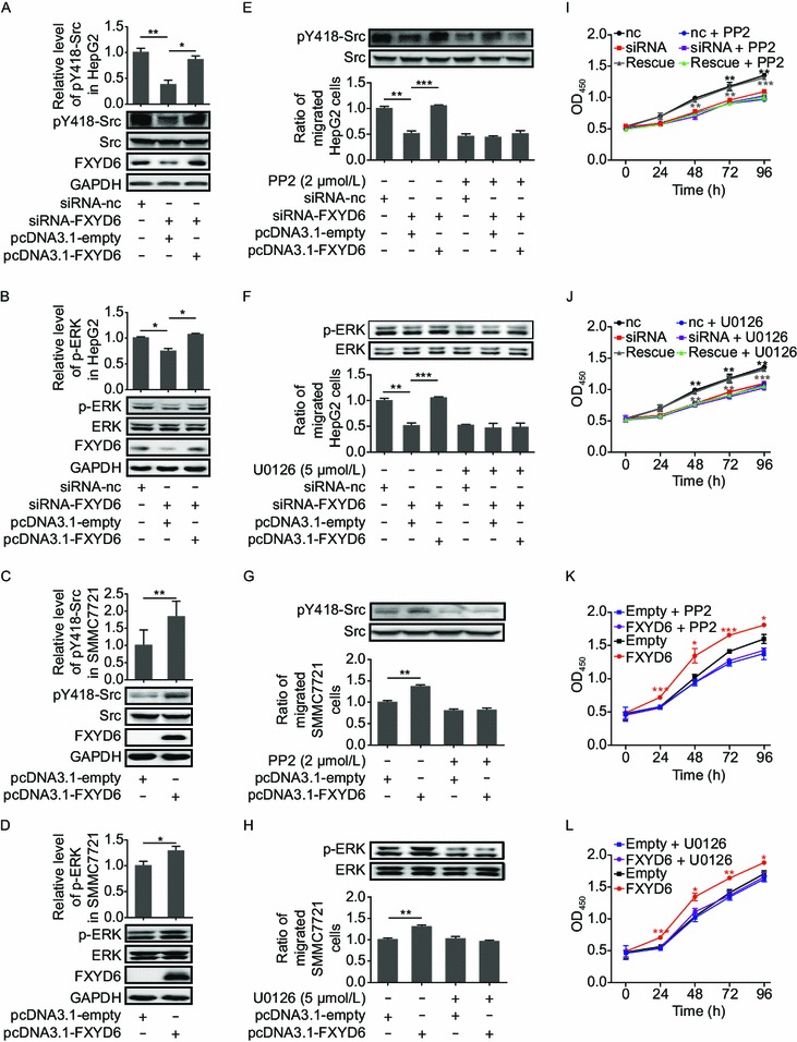
Figure 4.

FXYD6 activates Src and ERK signaling pathways in HCC cells. (A and B) The phosphorylations of Y418-Src (A) and ERK (B) were determined in HepG2 cells co-tranfected with siRNA-FXYD6 and pcDNA3.1-FXYD6. *P < 0.05 or **P < 0.01, compared with the siRNA-FXYD6 group. (C and D) The phosphorylations of Y418-Src (C) and ERK (D) were determined in SMMC7721 cells transfected with pcDNA3.1-FXYD6. *P < 0.05 or **P < 0.01, compared with the pcDNA3.1-empty group. The band density (mean ± SEM) was measured from at least 3 independent immunoblots and was normalized to the paired total. The signal from control cells was put to one. The representative images are shown. (E and F) The phosphorylation of Y418-Src in the presence of Src inhibitor PP2 (2 μmol/L) (E, upper panel) or the phosphorylation of ERK in presence of ERK inhibitor U0126 (5 μmol/L) (F, upper panel) was determined by immunoblotting and the migration (lower panel) of HepG2 cells was determined after co-transfection with siRNA-FXYD6 and pcDNA3.1-FXYD6. **P < 0.01 or ***P < 0.001, compared with the siRNA-FXYD6 alone group. (G and H) The phosphorylation of Y418-Src in the presence of Src inhibitor PP2 (2 μmol/L) (G, upper panel) or the phosphorylation of ERK in the presence of ERK inhibitor U0126 (5 μmol/L) (H, upper panel) was determined by immunoblotting and the migration (lower panel) was determined in SMMC7721 cells transfected with pcDNA3.1-FXYD6. **P < 0.01, compared with the pcDNA3.1-empty alone group. (I and J) The proliferation of HepG2 cells was determined after co-transfection with siRNA-FXYD6 and pcDNA3.1-FXYD6 in the presence of Src inhibitor PP2 (2 μmol/L) (I) or ERK inhibitor U0126 (5 μmol/L) (J) at different time points. **P < 0.01 or ***P < 0.001, compared with the siRNA-FXYD6 alone group. Rescue assay was conducted in HepG2 cells co-transfected with siRNA-FXYD6 and pcDNA3.1-FXYD6. (K and L) The proliferation was determined in SMMC7721 cells transfected with pcDNA3.1-FXYD6 in the presence of Src inhibitor PP2 (2 μmol/L) (K) or ERK inhibitor U0126 (5 μmol/L) (L) at different time points. *P < 0.05, **P < 0.01, or ***P < 0.001, compared with the pcDNA3.1-empty alone group. Data were collected from 3 independent experiments and the number of migrated cells from the controls was normalized as one
