
Fig 2.
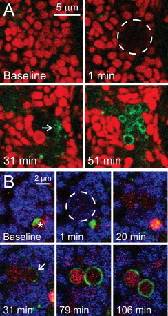
Type 2 mitophagy after selective photodamage. In (A), a TMRM-loaded GFP-LC3 hepatocyte was exposed to photodamaging 488-nm laser light within the area indicated by the circle. Note mitochondrial depolarization at 1 min after photoirradiation, followed by decoration of the depolarized mitochondria with GFP-LC3 (31 min, arrow). GFP-LC3 subsequently formed complete rings around the damaged mitochondria (51 min). In (B), a GFP-LC3 hepatocyte was loaded with ΔΨ-indicating MitoFluor Far Red (MFFR) and LysoTracker Red (LTR) for 30 min and then exposed to photodamaging 488-nm laser light within the area indicated by the circle. Note mitochondrial depolarization after photoirradiation, as indicated by loss of blue pseudo-colored MFFR fluorescence (1 min). GFP-LC3 subsequently began to decorate the depolarized mitochondria (31 min, arrow) to form mitophagosomes, which acidified as indicated by uptake of red LTR fluorescence (79 and 106 min). A pre-existing autophagosome was also present (baseline, asterisk), which matured into a red-fluorescing autolysosome and moved out of the field during the experiment.
Adapted from [25].