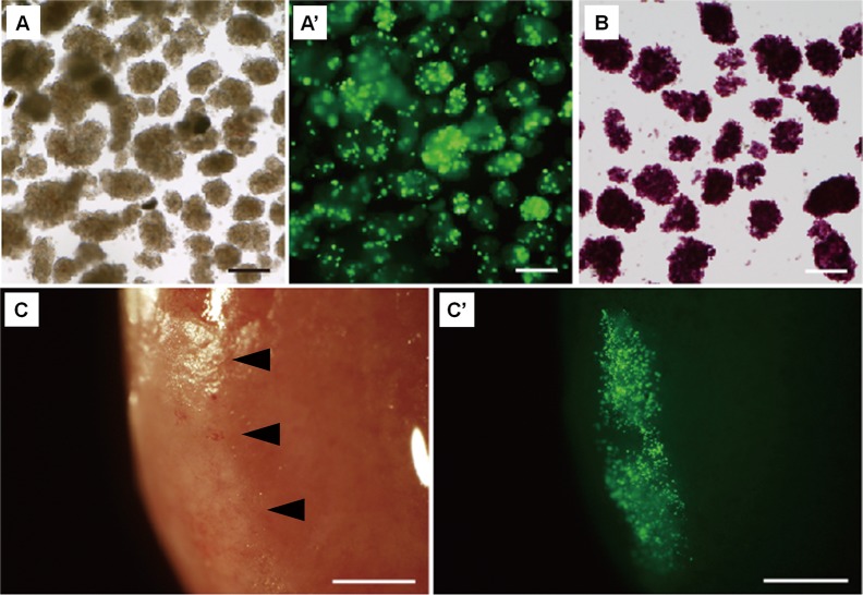
Fig. 4.
Fluorescence of pancreatic islets isolated from a Pdx1-Venus Tg pig. (A) Pancreatic islets isolated from a Tg pig. (A’) Fluorescent spots were observed in the islets of a Tg pig. (B) Dithizone-stained islets of a Tg pig. (C, C’) Pancreatic islets of a Pdx1-Venus Tg pig transplanted into the kidney capsule of NOD/SCID mice (arrowheads). Bright-field (C) and fluorescence (C’) observation by fluorescence stereomicroscopy showed that the fluorescence of the transplanted islets was clear at 30 days after transplantation (A’). Scale bars = 200 μm (A–C); 1 mm (C, C’).

