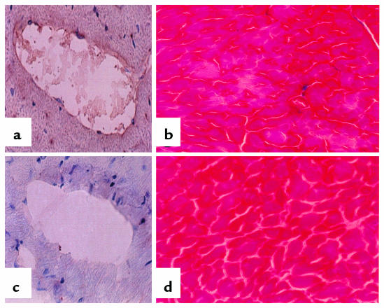
Figure 3.
Histology of heart. (a and c) Fibrin immunohistochemistry. Frozen tissue sections of tPA–/–TM–/Pro (a) and wild-type (c) mice were fixed in acidic formalin to wash out soluble fibrinogen/fibrin; reacted with the fibrinogen/fibrin-specific antibody; and counterstained with hematoxylin. The dark-brown horseradish peroxidase reaction product shows fibrin present in the macrovasculature. (b and d) Trichrome stain. Frozen tissue sections of tPA–/–TM–/Pro (b) and wild-type (d) mice were processed according to standard protocols (24). The tPA–/–TM–/Pro section shows myocardial infarct caused by the fibrin present in distal arteries. This tissue death is not present in the wild-type hearts. All images are sections from the LV wall of the heart. ×150.

