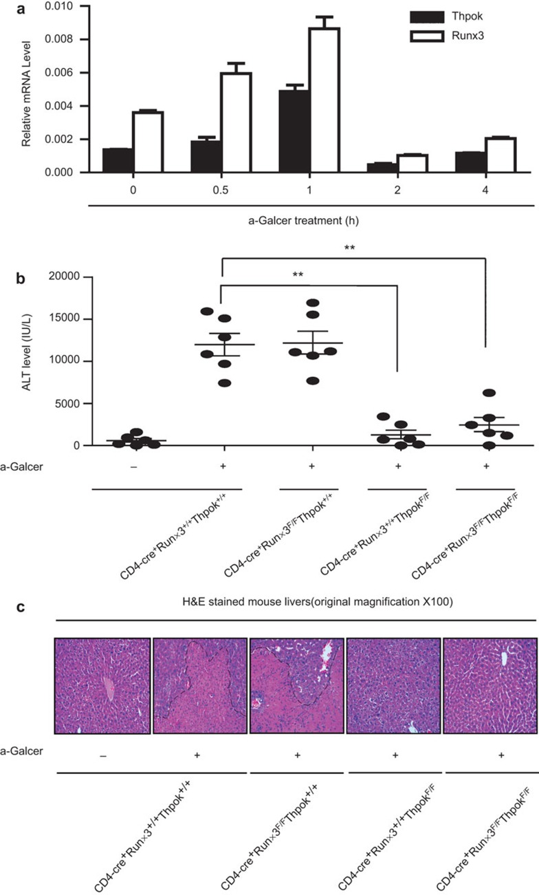
Figure 3.
Assessment of hepatic injury in Runx3-deficient, ThPok-deficient and DKO mice. (a) mRNA expression of Runx3, ThPok and Runx1 after αGalCer treatment at the indicated time points in WT mice. Mice were injected with 40 µg/kg of αGalCer, and liver MNCs and total cellular RNA were prepared at different time points. Runx3, ThPok and Runx1 levels were assayed by RT-PCR. Expression of each gene was normalized to β-actin expression. Data are representative of more than three independent experiments. (b) Serum ALT levels in mice lacking Runx3 or ThPok or both and littermate controls were measured 24 h after αGalCer treatment (or no treatment) and presented as the mean±s.e.m.. Each plot represents an individual mouse (n=6); ***P<0.001. (c) Runx3-deficient, ThPok-deficient, DKO and WT mice were challenged with αGalCer or vehicle for 24 h. Livers were then removed, sectioned and stained with H&E and evaluated by microscopy. The photomicrographs display the liver damage (original magnification: ×100) and are representative of at least three groups of separate experiments. αGalCer, α-galactosylceramide; ALT, aminotransferase; DKO, double knockout; H&E, hematoxylin and eosin; MNC, mononuclear cell; Runx3, runt-related transcription factor 3; WT, wild-type.

