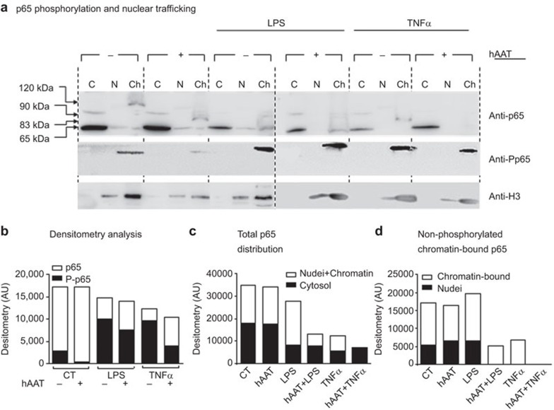
Figure 6.
hAAT promotes a distinct p65 phosphorylation and nuclear translocation pattern. RAW 264.7 cells (8×10⁶ per plate) were pre-treated with hAAT (0.5 mg/ml) for 1 h, washed and treated with LPS (10 ng/ml) or TNF-α (10 ng/ml). After 30 min, the cells were lysed and fractionated into cytosolic (C), nuclear (N) and chromatin-enriched fractions (Ch). The lysates were analyzed via western blotting with anti-p65 and anti-P-p65 antibodies. Tubulin and histone 3 were used as loading controls (tubulin not shown). (a) A representative blot from 10 repeats. (b) Densitometry analysis. (c) p65 distribution between the cytoplasm and nucleus. (d) Non-phosphorylated p65 distribution between all nuclear (the sum of nuclear membrane-associated p65, free nuclear p65 and chromatin-bound p65) and chromatin-enriched fractions. hAAT, alpha-1-antitrypsin; P-p65, phosphorylated p65.

