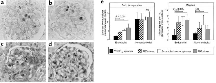
Figure 2.
Glomerular cell proliferation at 48 hours after induction of anti–Thy 1.1 nephritis in rats receiving the VEGF165 aptamer (n = 6), PEG alone (n = 6), scrambled VEGF165 aptamer (n = 5), or PBS alone (n = 5). (a) BrdU incorporation in a rat receiving VEGF165 aptamer. Labeled cells are mostly localized to the mesangium (cluster of 3 cells in the center of the glomerulus); only 1 glomerular endothelial cell (top edge of the glomerulus) is labeled. ×1,000. (b) BrdU incorporation in a rat receiving PEG alone. Labeled cells are mostly endothelial cells. ×1,000. (c) PAS-stained renal section from a rat receiving VEGF165 aptamer. A mesangial cell mitosis is present (arrow). ×1,000. (d) PAS-stained renal section of a rat receiving PEG alone. An endothelial cell mitosis is present (arrow). ×1,000. (e) Quantitative evaluation of endothelial vs. nonendothelial glomerular cell proliferation (as defined by counts of mitotic figures or nuclei incorporating BrdU). NS, not significant.

