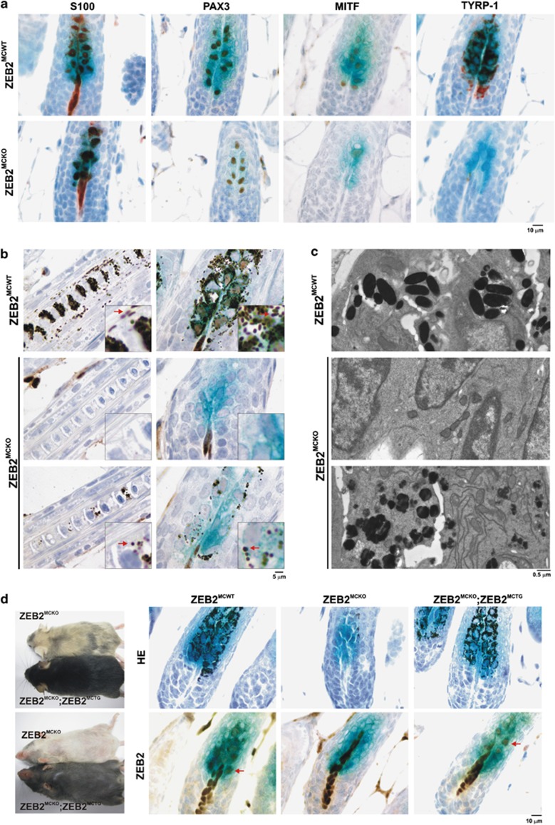
Figure 2.
Melanocyte-specific Zeb2 deficiency causes the formation of undifferentiated melanocytes in the bulge area of hair follicles. (a) Immunohistochemical staining of sections of ZEB2MCWT and ZEB2MCKO;Dct-LacZ-positive skin sections with S100b, PAX3, MITF and TYRP1 antibodies. (b) Detailed microscopic analysis of melanosomes in the hair shafts and the bulb area of the hair follicles, combined with immunohistochemical staining of ZEB2 on LacZ-positive skin sections of ZEB2MCWT and ZEB2MCKO; Dct-LacZ-positive mice. Insets show the altered morphology of melanosomes in the ZEB2MCKO sections compared with ZEB2MCWT. (c) Electron microscopic analysis of melanosomes in the bulb area of ZEB2MCWT and ZEB2MCKO mice demonstrates the absence or irregular morphology of melanosomes in the ZEB2MCKO hair follicles. (d) Genetic compensation of the loss of Zeb2 in the ZEB2MCKO;Dct-LacZ mice with melanocyte-specific overexpression of ZEB2 (ZEB2MCTG). Left panel: complete compensation of pigmentation in ZEB2MCKO ZEB2MCTG;Dct-LacZ mice compared with ZEB2MCKO;Dct-LacZ mice. Right panels: reappearance of pigmented melanosomes and ZEB2 expression in the ZEB2MCKO ZEB2MCTG;Dct-LacZ hair follicles. All microscopic analyses were done on skin sections of 5.5-day-old (a–c) or 13.5-day-old mice (d) and Immunohistochemical micrograph images were taken with a × 60/0.8 objective (a and d) or a × 100/1.25 objective (b)

