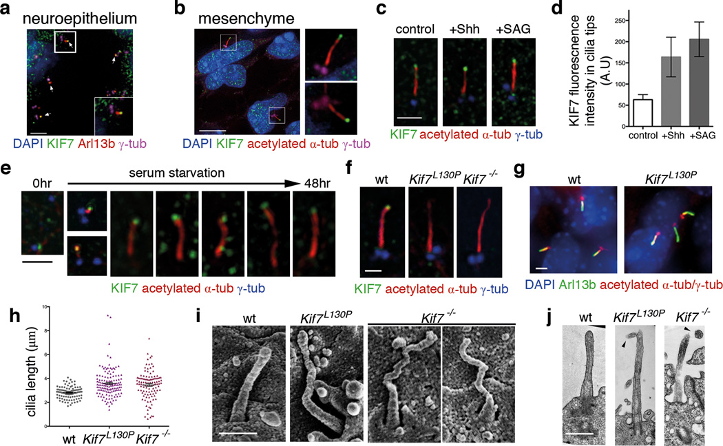
Figure 1. KIF7 constrains the length of primary cilia.
(a–b) KIF7 (green) localizes to the tips of primary cilia in e10.5 wild-type embryos. Arl13b (red) marks cilia in (a) and acetylated-α-tubulin (red) marks microtubule axoneme in (b). γ-tubulin (magenta) marks basal bodies. DAPI (blue) labels nuclei. Scale bar = 5µm. (c) In wild-type MEFs, KIF7 (green) is enriched in tips of primary cilia upon treatment with Shh-N recombinant protein (2µg/ml) or SAG (100nM) for 24 hours. Scale bar = 2µm. (d) Quantitation of KIF7 fluorescence intensity shown in (C). n = 150 cilia pooled from 3 independent experiments were counted for each condition. The error bars represent the SD. p<0.0001 between control and SAG or Shh treatments by one-way ANOVA. (e) KIF7 (green) is associated with one of the two centrioles prior to ciliogenesis and localizes to the tips of primary cilia throughout cilia elongation induced by serum starvation in wild-type MEFs. (f) KIF7 is not detected at the tips of Kif7L130P or Kif7−/− MEF cilia. Acetylated-α-tubulin (red) marks primary cilia and γ-tubulin (blue) marks basal bodies in (c), (e) and (f). Scale bar = 1µm in (c), (e) and (f). (g) Primary cilia of wild-type and Kif7L130P MEFs stained with Arl13b (green), acetylated-α-tubulin and γ-tubulin (red). DAPI in blue. Scale bar = 2µm. (h) Measurements of MEF cilia length between wild-type and Kif7 mutants using Arl13b as cilia marker. >100 cilia pooled from 4 independent experiments were counted for each genotype (n=108 cilia for wild type, n=133 cilia for Kif7L130P mutant, and n=111 for Kif7−/− mutant). p<0.0001 between wild-type cilia and Kif7 mutant cilia by one-way ANOVA. The error bars represent the SEM. (i) Scanning electron microscopy shows neural tube cilia of e10.5 wild-type, Kif7L130P and Kif7−/− embryos. Wild-type neural cilia were 0.97±0.17 µm and Kif7L130P neural cilia were approximately 1.2±0.28µm long (n=50 for each genotype). Scale bar = 0.5µm. (j) TEM images of longitudinal sections of wild-type, Kif7L130P and Kif7−/− neural tube cilia. Arrowheads point to twisted tips of mutant cilia. Scale bar = 0.5µm.

