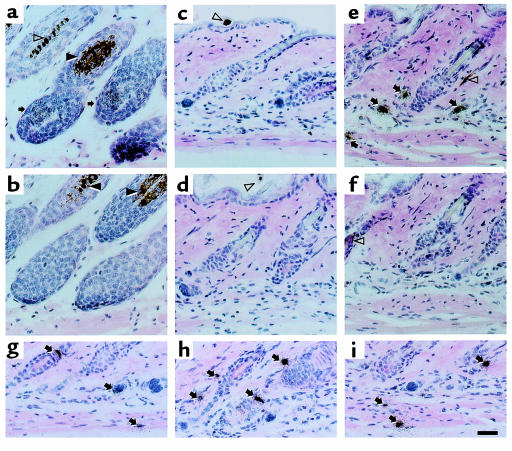
Figure 2.
Localization of Shh expression in skin by in situ hybridization before and after administration of AdShh. Paraffin sections of mouse skin during its natural anagen period (postnatal day 33) compared with postnatal day 22 naive mouse skin or skin of mice injected on postnatal day 19 with PBS, AdNull, or AdShh. The sections were analyzed by in situ hybridization using [33P]UTP-labeled antisense and sense Shh riboprobes. After hybridization, sites of probe binding were identified using a photographic emulsion, and tissue was stained with hematoxylin and eosin. Arrows indicate positive staining for Shh mRNA. Closed arrowheads indicate melanosomes in the hair follicle. Open arrowheads indicate melanin in hair shafts. (a) Anagen skin from a naive 33-day-old mouse (positive control) hybridized with an antisense Shh complementary to Shh mRNA. (b) Adjacent tissue section of anagen skin hybridized with a sense Shh probe. (c) Naive postnatal day 22 skin hybridized with an antisense Shh probe. (d) Postnatal day 22 skin 3 days after injection with 108 PFU of AdNull hybridized with an antisense Shh probe. (e) Postnatal day 22 skin 3 days after injection of 108 PFU of AdShh hybridized with an antisense Shh probe. (f) Postnatal day 22 skin 3 days after injection with 108 PFU of AdShh hybridized with a sense Shh probe. (g–i) High-magnification examples of cells in AdShh-injected mouse skin hybridized with antisense Shh probe. Scale bar: 50 μm.

