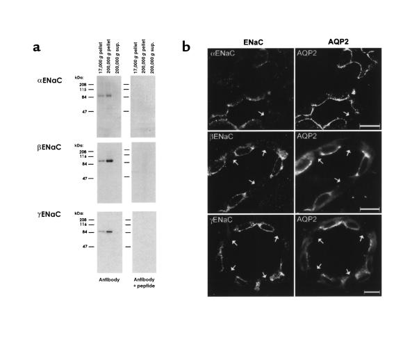
Figure 1.
Characterization of ENaC subunit antibodies. (a) Immunoblots of rat renal cortical proteins obtained by differential centrifugation. Blots were probed with the 3 ENaC subunit antibodies and with the antibodies preadsorbed with 1 mg of the respective immunizing peptides. Differential centrifugation was carried out as described (6), yielding membrane fractions (17,000 g and 200,000 g pellets) and a cytosolic fraction (200,000 g supernatant). (b) Immunofluorescence localization of ENaC subunits in rat renal cortical collecting duct. Three sections are shown, each double-labeled with 1 of 3 rabbit antibodies to ENaC subunits (left-hand panels) and a chicken anti–aquaporin-2 (right-hand panels). Arrows point to cells lacking aquaporin-2, i.e., intercalated cells. All tissue sections were from sodium-restricted rats. Scale bars: 10 μm.

