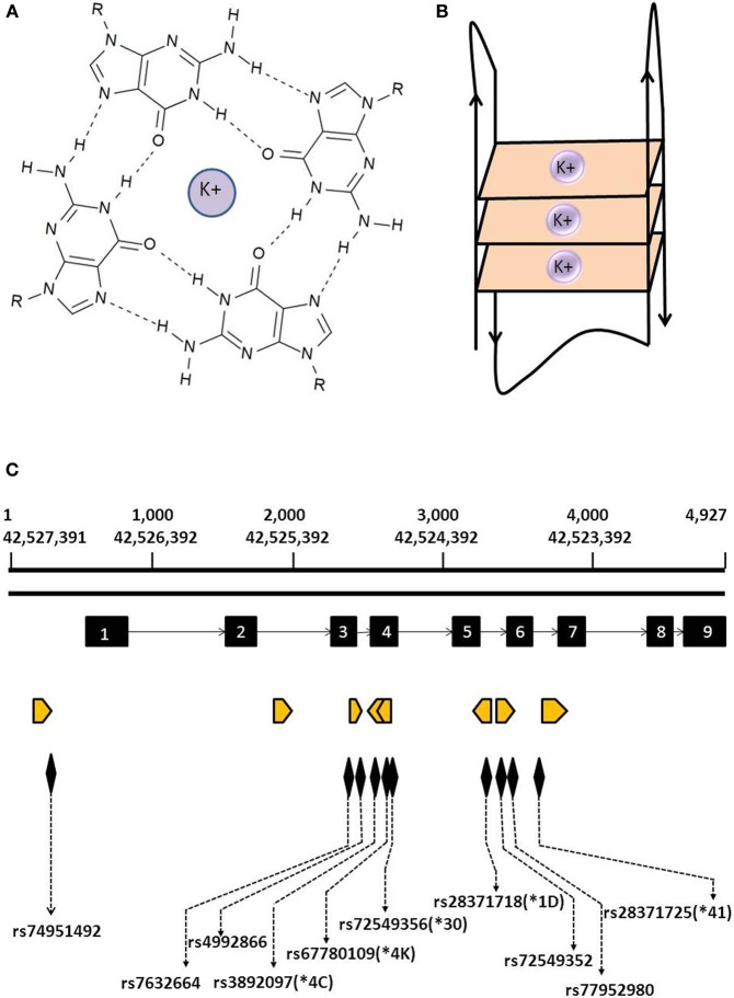
Figure 1.
Structure of G4 and location of predicted G4s within the CYP2D6 gene. (A) Guanine tetrad formed by the association of four guanine bases via Hoogsten hydrogen bonds in a coplanar arrangement and stabilized by a potassium ion. (B) Schematic representation of a G-quadruplex formed by the stacking of the three guanine tetrads from a single strand of DNA. (C) Location of predicted G4s in the CYP2D6 gene. Orange arrowheads indicate location and orientation of predicted G4s; exons are depicted by black blocks. Also indicated are the locations of SNPs and CYP2D6* alleles found within predicted G4s.

