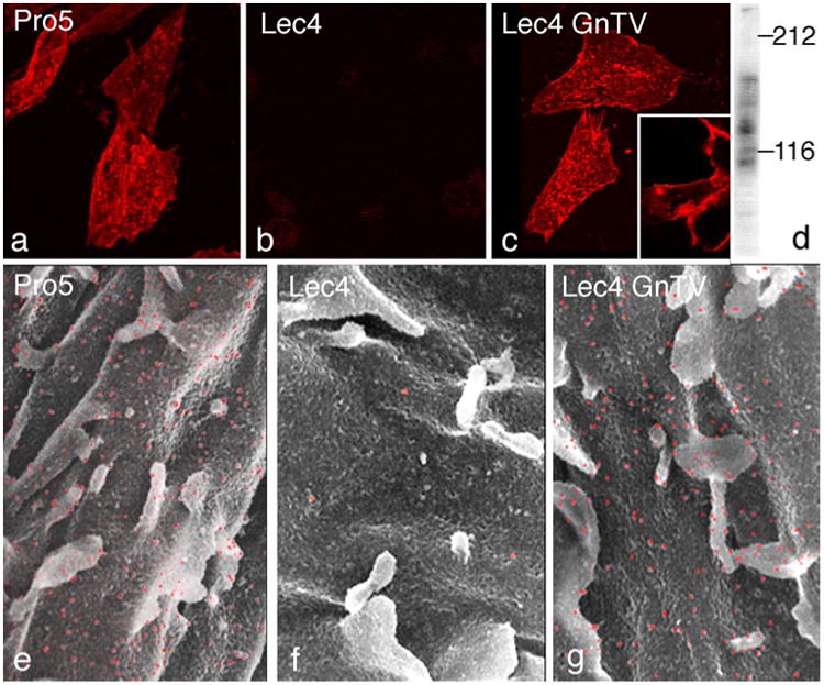
Fig. 1.

Demonstration of β1,6-branched N-glycans on the CHO cell surface using dig L-PHA and confocal laser scanning microscopy (a–c) and lectin-gold scanning electron microscopy (e–g). The CHO Pro−5 cells (a, e) and CHO Lec4 GnTV-N5 cells (c, g) showed positive cell surface staining with microvilli being strongly positive. In contrast, CHO Lec4 cells (b, f) showed no specific cell surface labeling. ×1,100 (a–c), ×9,000 (e–g). Dig L-PHA—blot of homogenates from CHO Lec4 GnTV cells revealed two major bands at 140 and 85 kDa and several minor bands (d)
