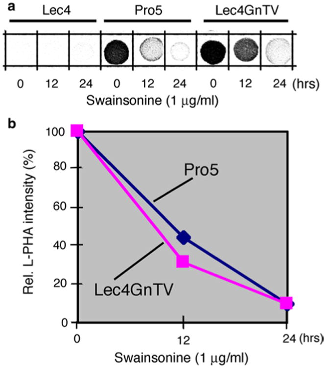
Fig. 5.

Reduction in the β1,6-branched N-glycans in CHO cells after swainsonine treatment. Swainsonine (1 μg/ml) was added to the cell culture medium, and cells were collected after 0, 12, and 24 h of treatment. Homogenates were processed for L-PHA spot blots, and the results densitometrically analyzed. The CHO Lec4 cells served as control
