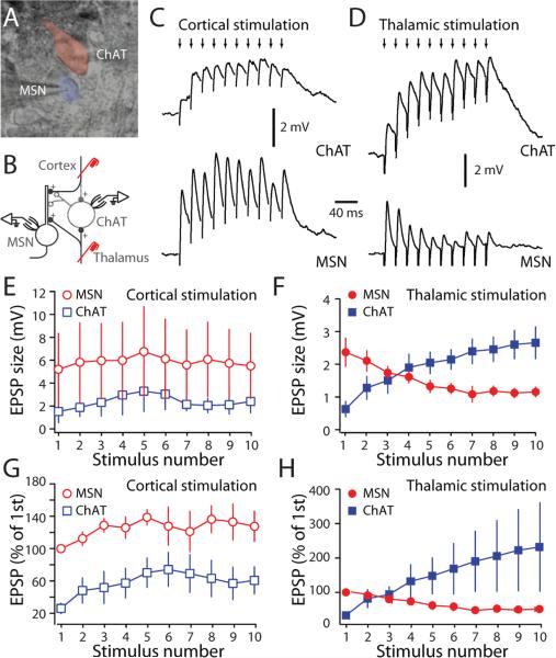
Figure 3. Thalamic stimulation elicited distinctive postsynaptic responses in interneurons and MSNs.
(A) Infrared differential interference contrast image showing an MSN and a cholinergic interneuron. (B) Schematic of the experimental configurations. (C) Paired recording from a cholinergic interneuron (ChAT) and a neighboring MSN during cortical stimulation. Cortical stimulation (small arrows, 50 Hz) evoked EPSPs in both cell types. However, EPSPs recorded in MSNs had a bigger amplitude. (D) Paired recording from ChAT) and neighboring MSN in response to train of thalamic stimulation. Thalamic stimulation (small arrows, 50 Hz) evoked EPSPs that summed differently in cholinergic interneurons and MSNs. (E) The evoked cortical EPSPs amplitudes in MSNs and cholinergic interneurons were plotted against stimulus number. (F) The evoked thalamic EPSPs amplitude in MSNs and cholinergic interneurons were plotted against stimulus number. (G) Normalized cortical EPSPs amplitude was plotted against stimulus number. Cortical EPSPs in cholinergic interneurons are smaller than those in MSNs. (H) Normalized thalamic EPSPs amplitude was plotted against stimulus number. Thalamic EPSPs in cholinergic interneurons summed differently than those in MSNs. To unmask EPSPs, hyperpolarizing current was injected into cholinergic interneuron to prevent cells from firing action potentials. Experiments were performed at 32-35°C.

