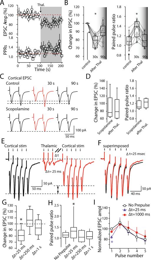
Figure 5. Corticostriatal synaptic transmission was depressed by thalamic stimulation.
(A) Time course of changes in cortical EPSC amplitude and PPRs in response to thalamic burst stimulation. Consistent with a presynaptic mechanism, there was a clear increase in PPRs accompanied by reduction in ESPC amplitude. (B) Box-plot summary of the change in first EPSC amplitude (left) and change in PPRs (right). (C) Sample EPSCs evoked by a paired-pulse cortical stimulation with 50 ms interstimulus interval. Traces illustrate the suppression of EPSC amplitude and increase in PPRs at cortical afferent synapses after burst stimulation of thalamic afferents. Scopolamine blocked the depression. (D) Box-plot summary of change in first EPSC amplitude (left) and changes in PPRs (right) in the presence of scopolamine. (E) Sample traces of cortical EPSCs recorded from a medium spiny neuron in the absence (left) and presence (right) of a thalamic pre-pulse with 25 ms interval (Δt=25 ms) at physiological temperature. (F) Cortical EPSCs were superimposed to illustrate the changes in EPSCs amplitudes (Δt=25 ms). (G) Box-plot summary of the reduction in first EPSC amplitude. Presynaptic modulation of cortical EPSCs by thalamic burst stimulation recovered quickly at higher temperature (Median amplitude=82% of control; P<0.05; median amplitude=108% of control at 250 ms, P>0.05, n=10;median amplitude=94% of control at 1s , P>0.05, ANOVA; n=10). (H) Box-plot summary of the paired pulse ratio (Mean control PPR=1.11; PPR(25ms)=1.30, P<0.05; PPR(250ms)=1.09, P>0.05, ;PPR(1s)=1.09, P>0.05, ANOVA; n=10). (I) Pooled data (n = 10) showing the effect of the thalamic pre-pulse on short term plasticity of EPSCs evoked by 50 Hz cortical afferent stimulation. Experiments illustrated in A-D were performed at room temperature; E-I were performed at 32-35°C.

