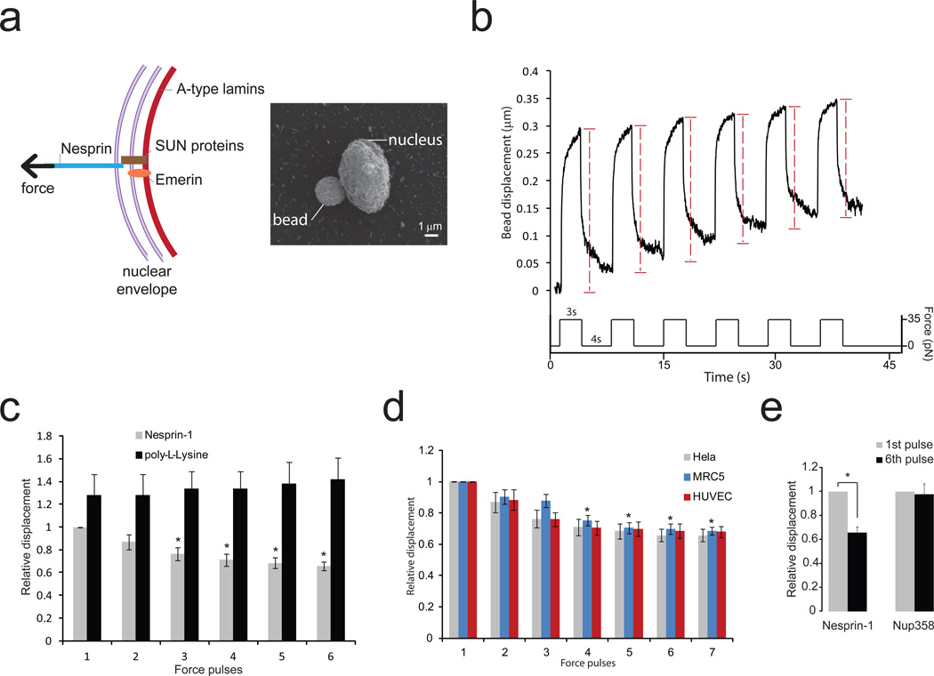
Figure 1. Isolated nuclei stiffen in response to force applied on nesprin-1.
a, Diagram of the LINC complex (left panel) showing where tensional forces were applied in order to mimic the transmission of mechanical stress from the cytoskeleton to the nucleus. Scanning electron microscope picture of a magnetic bead attached to a nucleus isolated from a Hela cell (right panel). Result is representative from 6 independent experiments.
b, Typical displacement of a 2.8 µm bead coated with anti nesprin-1 antibody bound to an isolated nucleus during force pulse application. Stiffening is indicated by decreased displacement during later pulses.
c, Change in bead displacement during 6 force pulses applied to beads coated with anti nesprin-1 antibody (n=18 beads) or poly-L-lysine (n=14 beads) and bound to nuclei isolated from Hela cells. Displacements were calculated relative to the first pulse of force applied to beads coated with anti nesprin-1 (Error bars represent s.e.m., *P<0.05, data were collected from 3 independent experiments and analyzed by one way ANOVA).
d, Change in bead displacement during 7 force pulses applied to beads coated with anti nesprin-1 bound to nuclei isolated from Hela cells (n=18 beads), MRC5 cells (n=21 beads) or HUVECs (n=15 beads). Displacements were calculated relative to the first pulse of force (Error bars represent s.e.m., *P<0.05, data were collected from 3 independent experiments and analyzed by one way ANOVA).
e, Change in bead displacement between the first and 6th pulse of force applied to beads coated with anti nesprin-1 antibody (n=18 beads) or anti NUP358 antibody (n=16 beads) and bound to nuclei isolated from Hela cells. Displacements were calculated relative to the first pulse of force (Error bars represent s.e.m., *P<0.05, data were collected from 3 independent experiments and analyzed by two-tailed unpaired t-test).

