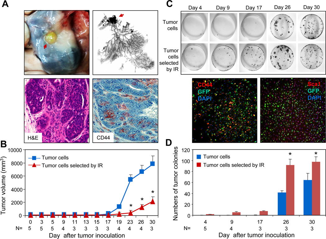
FIGURE 3.
Tumorigenic and metastatic potential of 6Gy-enriched tumor cells. (A-B) 1×10⁶ 6Gy-selected or unselected tumor cells isolated from GFPMMT mice were injected into the left and right mammary fat pads of syngeneic WT mice. The mice were sacrificed on the indicated day and the mammary glands harvested. The red arrow indicates the tumor in the mammary glands (A, upper panels). The mammary glands were further processed for sections that tumor were stained with H&E or anti-CD44 mAb and examined under microscope (60x). The red color indicates positive cells (A, lower right panel). (B) The growth of tumor cells that received 0 or 6Gy radiation was presented. The statistical significance was determined by t-test (p<0.001). (C) Colony-forming assay of lung cells from recipient mice. Lungs were harvested and perfused to remove the circulating cells. The lung tissue was minced and digested in collagenase enzyme cocktail solution. The cells were cultured in DMEM medium supplemented with 10% FCS for two weeks and stained with 0.5% Crystal Violet. In lower panel, a colony formed in the lung cell culture on day 30 from mice inoculated with tumor cells irradiated by ionizing radiation (IR) was stained with PE conjugated anti-CD44 (left) or Sca1 (right) mAbs and examined under fluorescent microscope (10x). (D) The numbers of tumor colonies from the lungs of recipient mice were counted and presented. The statistical significance was determined by t-test. (*) indicates p=0.006 on day 26 and p=0.013 on day 30).

