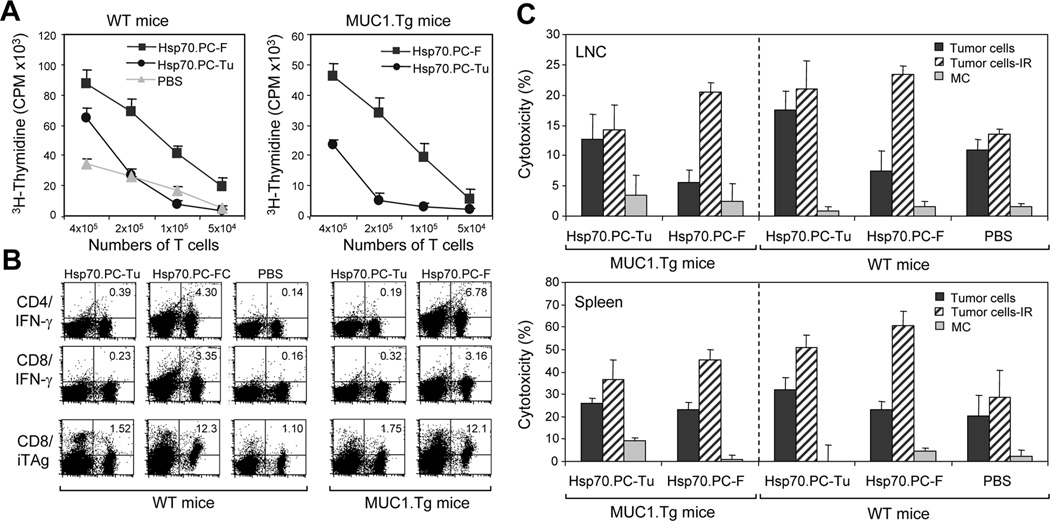
FIGURE 4.
T cell response elicited by HSP70.PC-F vaccine prepared from 6Gy-selected tumor cells. (A) T cell proliferation assay. Draining lymph node cells (LNC) obtained from WT mice (left panel) or MUC1.Tg mice (right panel) immunized with HSP70.PC derived for fusion cells of DC and 6Gy irradiated tumor cells (Hsp70.PC-F, ■) or 6Gy irradiated tumor cells (Hsp70.PC-Tu, ●). Control mice were injected with PBS (▲). LNC were cultured for 5 days and [3H]-thymidine was added in the last 12 hours of culture. The incorporation of [3H]-thymidine was measured. (B) LNC obtained from immunized mice were analyzed by FACS for expression of IFN-γ in CD4 and CD8 T cells and/or MUC1 tetramer in CD8 T cells. The percentage of double positive cells were indicated. (C) CTL assay. LNC and splenocytes were isolated from mice twice immunized with Hsp70.PC-F, Hsp70.PC-Tu or PBS, and incubated with 51Cr-labeled ionizing radiation (IR) selected tumor cells, nonirradiated tumor cells or monocytes (MC) at 60:1 (LNC) or 100:1 (splenocytes) E: T ratios. CTL activity was determined by 51Cr-release assay.

