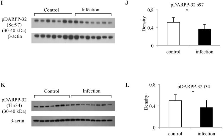
Figure 4. Reduced expression of several proteins involved in dopamine pathway in mouse striatum at day 5 postinfection.
Protein expression of (A) DRD1; (C) DRD5; (E) MAOA; (I) DARPP-32 Phosphorylation at Ser97; and (K) DARPP-32 Phosphorylation at Thr34 between control and infected mice. Quantification of normalized level of (B) DRD1; (D) DRD5; (F) MAOA; (J) DARPP-32 Phosphorylation at Ser97; and (L) DARPP-32 Phosphorylation at Thr34 between control and infected mice. (G) Total DARPP-32 expression is comparable between control and infected mice. (H) Quantification of normalized level of DARPP-32 shown in (G). Protein levels were compared to β-actin. Control mice: n = 6 - 7; Infected mice: n = 6 - 8; Error bars = 1 SD. * < 0.05, ** < 0.01.


