
Fig. 3. PTH 1-34 enhances bone formation, but not cartilage formation, following tibia fracture in both young and aged mice.
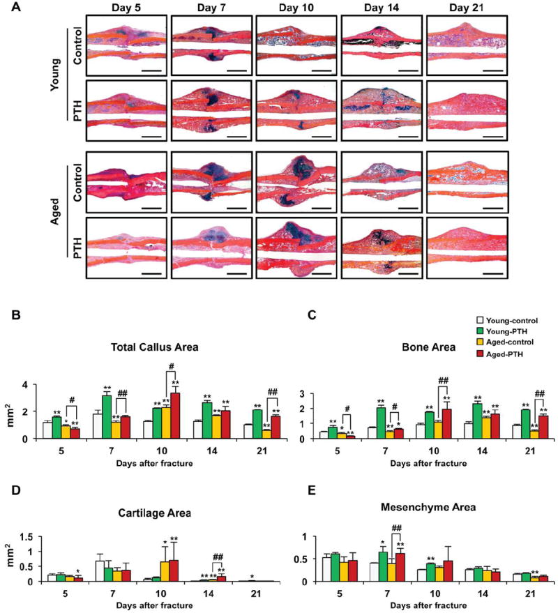
(A) Representative Alcian blue hematoxylin/orange G stained sections from the fractures of young and aged mice treated with saline or PTH 1-34 for 5, 7, 10, 14, or 21 days. PTH 1-34 increased intramembranous bone formation in both young and aged mice, but delayed completion of endochondral bone formation evident by the presence of persistent cartilage at days 10 and 14 in PTH 1-34 treated fractures. Histomorphometry was used to measure total callus area (B), bone area (C), cartilage area (D), and mesenchyme area (E) in the fractures of young (n=4) and aged (n=4) mice treated with saline or PTH 1-34 for 5, 7, 10, 14, or 21 days. Three levels were measured per sample. PTH 1-34 increased total callus, mesenchyme, and bone area, but not cartilage area. Data are presented as mean ± standard error (SEM). Statistical analysis was performed using bootstrap-adjusted t-tests and statistical significance denoted as follows: *p<0.05 and **p<0.01 compared to Young control group, or #p<0.05 and ##p<0.01 for Aged control vs. Aged PTH 1-34 groups.