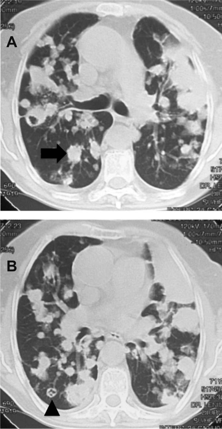
Fig. 1. A computed tomography scan revealing multiple pulmonary nodules (arrow in A) in the lower lobes. The scan also demonstrates some cavitary lung lesions (arrowhead in B) with feeding vessel sign in the lower lobes. These imaging findings suggest secondary vascular implants that could be of infectious or neoplastic etiology.

