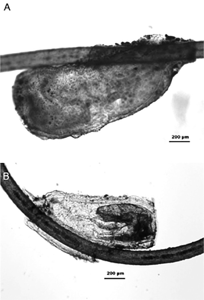
Fig. 1. Eggs/nits under bright field microscopy. After the rehydration process, it is possible to visualize the embryonic stage inside the eggshell. 50 nits were measured: the size ranged between 1,126.92 µm (length) and 469.38 µm (width). Scale bar = 200 µm.

