. 2014 May-Jun;56(3):259–264. doi: 10.1590/S0036-46652014000300012

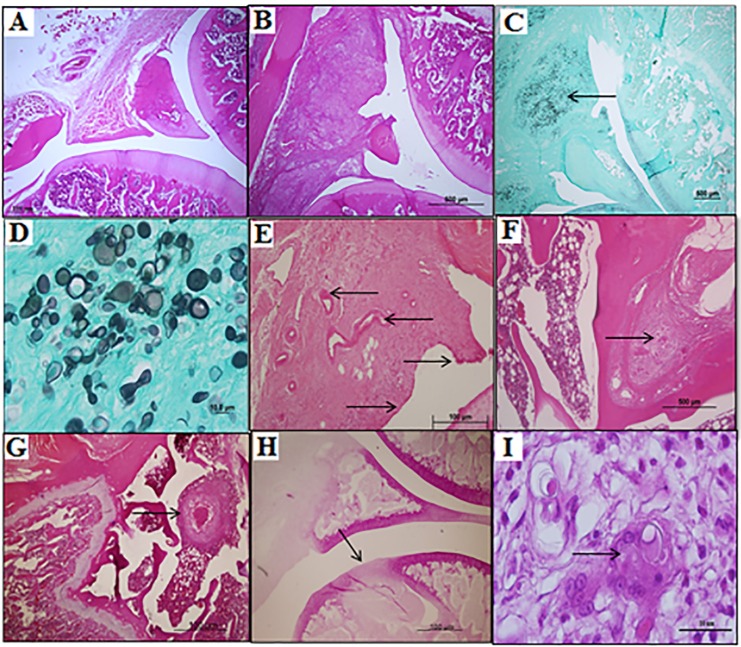
Fig. 2. Photomicrographs of histological longitudinal sections from the knees of the experimental animals. A - Articulation of a normal animal, GCA (HE 4x). B - Synovial membrane with swelling and granuloma from an animal after 15 days of exposure to a dose of 103 yeast cells (HE 4x). C - Synovial swelling with a large amount of P. brasiliensis cells, indicated by the arrow, from an animal exposed to a dose of 104 yeast cells for 15 days (Grocott 4x). D - Detail of figure (C) with a large amount of P. brasiliensis cells with budding (Grocott 40x). E - Pannus formation with the emergence of vessels (left arrow) and the multilayered synovial mesothelium (the arrows to the right) at 45 days exposure (HE 10x). F - Granuloma with a giant cell bone inside (HE 10x). G - Granuloma bone above the epiphyseal line, focusing on the necrosis inside, in one animal after 45 days of exposure to 105 viable yeast cells (HE 4x). H - The arrow indicates the area of cartilage destruction, with a deep necrotic area after 45 days of exposure to 105 viable yeast cells (HE 4x). I - Detail of figure (F) with a giant cell with P. brasiliensis cells inside (HE 100x).

Fig. 2