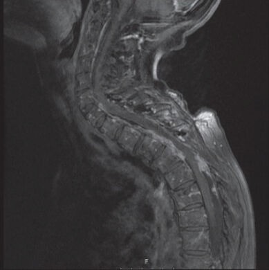
Figure 1.

Magnetic resonance imaging, T1 sequences with gadolinium, sagittal view. Imaging after two previous resections. Note strong dorsal (C7/Th1 to Th5) and ventral (C6 to Th3/Th4) enhancement in the spinal canal. Artifacts due to laminoplasty material
