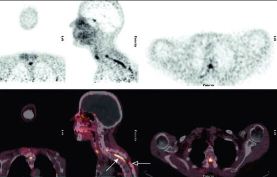
Figure 2.

68Ga-DOTATATE-positron emission tomography/computed tomography imaging. Coronal, sagittal and axial views. Images presented a high tracer uptake in the ventral part of the spinal canal esp. at the level C6-Th4 (solid arrow) as well as in the dorsal part at the level Th4 + 5 (hollow arrow) corresponding histological to vital tumor. Note typical intracranial enhancement of the pituitary gland
