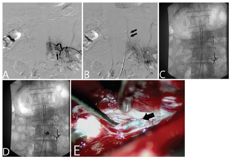
Fig. 3. Case 3.

A and B: Left L-3 segmental artery injection on spinal angiography (anteroposterior projection) in early arterial (A) and late arterial (B) phases revealing a DAVF fed by a radicular branch at this level (single arrow) with drainage into a slow flowing vein (double arrows). C and D: Left L-2 segmental artery injection on spinal angiography (anteroposterior projection) in early arterial (C) and late arterial (D) phases, demonstrating DAVF recurrence from a collateralized radicular branch under the L-3 pedicle with similar drainage into a slow flowing vein (double arrows). E: Visual inspection during surgical ligation revealing the presence of Onyx cast material within the draining vein (arrow) of the fistula.
