
Figure 2.
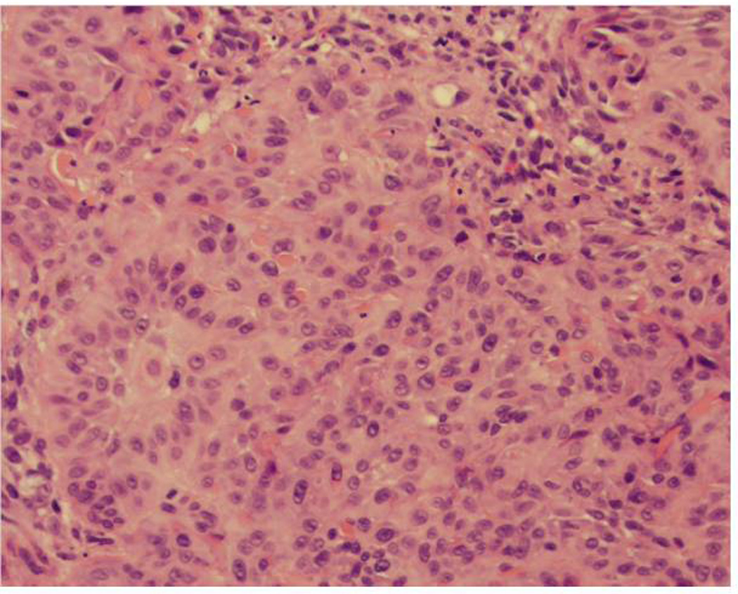
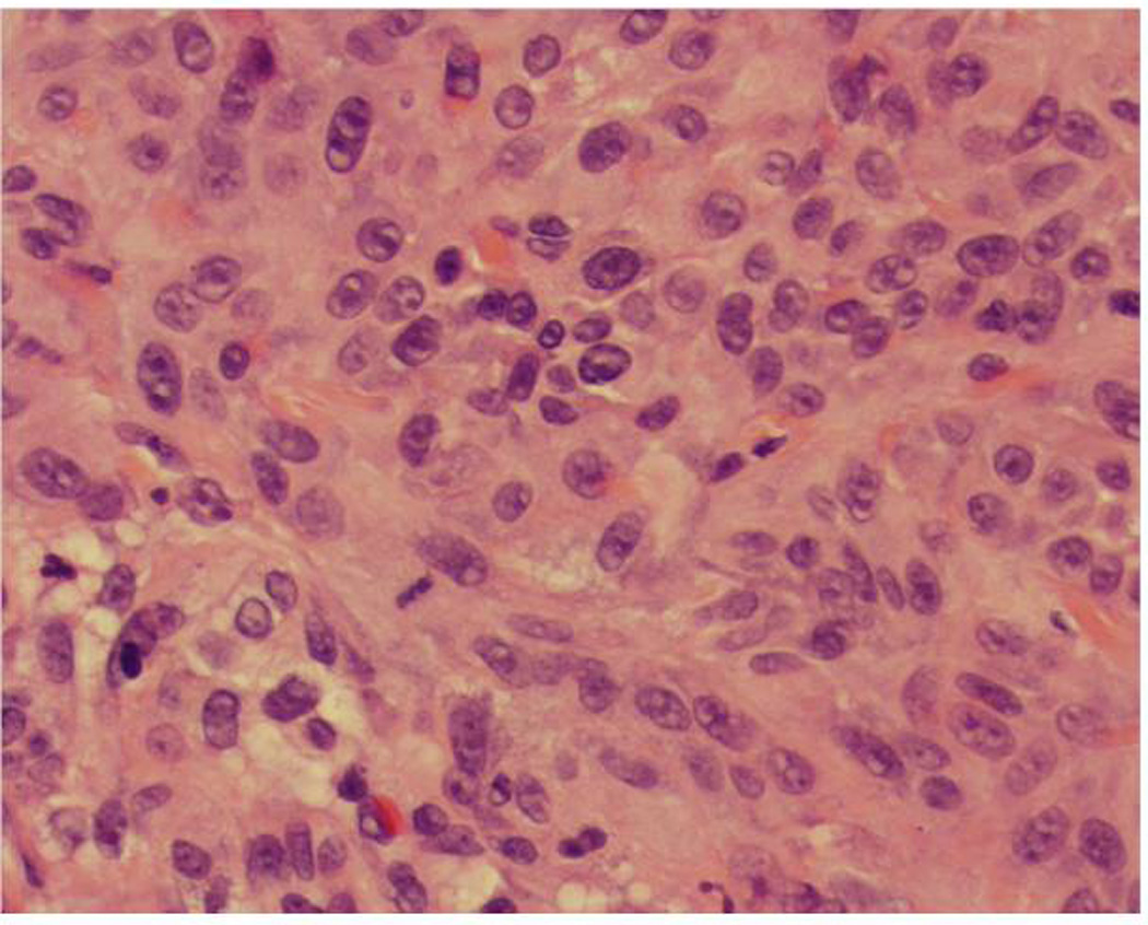
Invasive squamous cell carcinoma, moderately differentiated, 3.3 mm in thickness
(A) Marked cytologic atypia at the interface between the tumor and the dermis (×20)
(B) Notice the presence of large nuclei, prominent nucleoli and mitotic figures (×40)