
Figure 2.
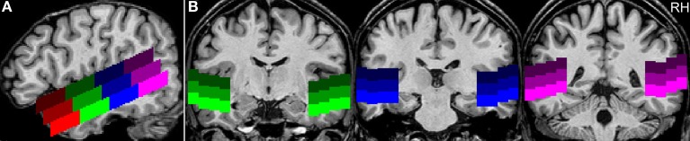
Example for the positioning of the regions of interest. (A) The sagittal view of a single subject's brain shows the position of the ROIs along the superior temporal sulcus at Talairach coordinate x = 50 (RH, right hemisphere). The upper row of ROIs covers the superior part of the STG (sSTG), the middle row the inferior part of the STG (iSTG), and the lower row the superior part of the MTG (sMTG). Red ROIs are located in the anterior temporal lobe (a), green ROIs in the mid-anterior part (ma), blue ROIs in the mid-posterior part (mp), and purple ROIs in the posterior temporal lobe (p). (B) Coronal views at y = −10 (green), −25 (blue), and −41 (purple).