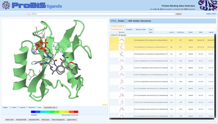
Figure 2.

ProBiS-ligands output page. Left: query protein (green cartoon model) and two predicted ligands (CPK colored stick models). Invariant binding sites residues are thinner CPK-colored sticks. Right: table with predicted small-molecule ligands clustered according to their predicted location on the query protein and transposed from different binding sites; the two selected ligands are highlighted.
