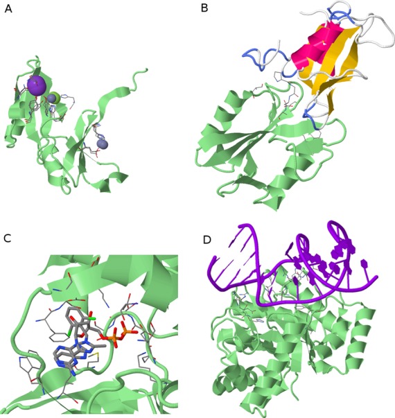
Figure 3.

Predicted protein–ligand complexes. Query proteins are green cartoon models and invariant binding site residues are CPK-colored stick models (Ligand View 1). (A) Three predicted ion ligand clusters (ions are spheres) on Glyoxalase family protein (PDB ID: 2qqz). (B) Predicted protein ligand (yellow–pink cartoon) on SH2 domain protein (3tkz). (C) Two predicted small molecule ligands, i.e. ATP and an inhibitor of biotin carboxylase (thick CPK sticks) on D-alanine:D-alanine ligase (1iov). (D) Predicted DNA ligand on endonuclease IV protein (4hno).
