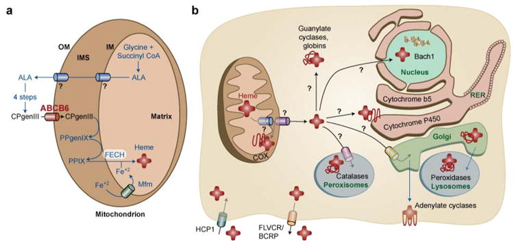
Fig. 1.

Schematic model of mammalian heme homeostasis. Presumptive heme pathways that are currently unknown are marked with a ? .
(A) In eukaryotic cells, heme is synthesized via a multi-step pathway that is spatially separated between the cytosol and mitochondria. The export of delta-aminolevulinic acid (ALA) from the mitochondria is unknown. ALA is converted to CPgenIII via four enzymatic reactions and CPgenIII is imported into the mitochondria by the outer membrane (OM) transporter, presumably ABCB6 or OGC. CPgenIII is converted to protoporphyrinogen (PPgenIX) and PPIX on the intermembrane space (IMS) side of the inner membrane (IM). The final step of heme synthesis occurs by the enzymatic chelation of ferrous iron (Fe+2) into PPIX catalyzed by ferrochelatase (FECH) located on the matrix side of the IM. Mitoferrin (Mfrn) / Mrs 3/4 is predicted to import Fe+2 into the mitochondria.
(B) The nascent heme moiety is somehow transported through two mitochondrial membranes and incorporated into a multitude of hemoproteins, presumably by hemochaperones, in different cellular compartments. Recent studies have identified HCP-1 as the heme importer, primarily in intestinal cells, while FLVCR and BCRP is predicted to export heme in erythrocytes.
