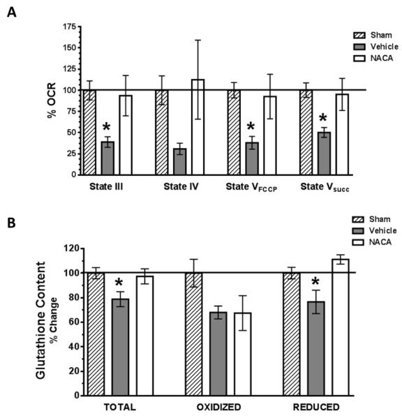
Fig. 3. NACA significantly improves mitochondrial oxidative phosphorylation and glutathione content following TBI.
(A). Cortical mitochondria were isolated at 25 hrs post-injury from sham operated or TBI injured animals that were either administered vehicle or NACA (150mg/kg) at 5 minutes and then every 6 hours up to 24 hrs post-injury. Mitochondrial respiration was then measured and oxygen consumption rates (OCR) calculated and expressed as % of sham animal values (%OCR). NADH-linked mitochondrial ATP synthesis (State III), Oligomycin (State IV), FCCP (State VFCCP) and FADH-linked (State Vsucc) dependent mitochondrial oxygen consumption rates were measured. Following TBI, a significant reduction (60-80%) in mitochondrial State III, State VFCCP and State Vsucc respiration was measured in vehicle treated animals. NACA treatment significantly improved all mitochondrial respiratory parameters as compared to vehicle treated animals following TBI, reaching the level of sham operated animals. (B). Cortical mitochondria were isolated at 25 hrs post-injury from sham operated or TBI injured animals using the same treatment paradigm as that used for mitochondrial assessments. Mitochondrial glutathione (GSH) content (total, oxidized and reduced form of GSH) was measured and expressed as % of sham operated levels. Following TBI there is a significant reduction (21-23%) in mitochondrial total and reduced GSH in vehicle as compared to sham or NACA treated animals. Bars represent group means, ± s.e.m. (n = 5 animals/group). Statistical analysis performed was an ANOVA followed by Fisher post hoc test. *p < 0.05 compared to sham or NACA treated group.

