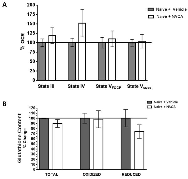
Fig. 4. NACA treatment show identical mitochondrial respiration rates and glutathione content compared to vehicle treated naïve rats.
Uninjured, experimentally naïve animals were injected with NACA (150mg/kg) or vehicle every 6 hrs for a duration of 24 hrs (5 injections). Mitochondrial function (A). and GSH levels (B). were then measured in the cortex. No significant differences in any parameter of mitochondrial bioenergetics or GSH levels were measured. Bars represent group means, ± s.e.m. (n = 3 animals/group). Statistical analysis was performed using unpaired t-tests.

