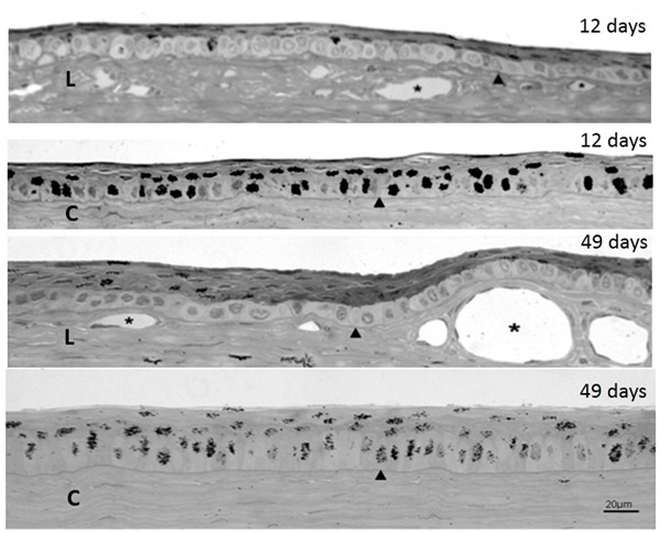
Figure 3. Autoradiography of semithin sections of rabbit eyes injected three times with [3H]-TdR and killed at 12 and 49 days after the first injection, showing both the limbus (L) and the center of the cornea (C). The cuboid clear cells making up the basal stratum in the limbus are conspicuous. Arrowheads indicate the basal membranes; asterisks are in the lumen of blood vessels.

