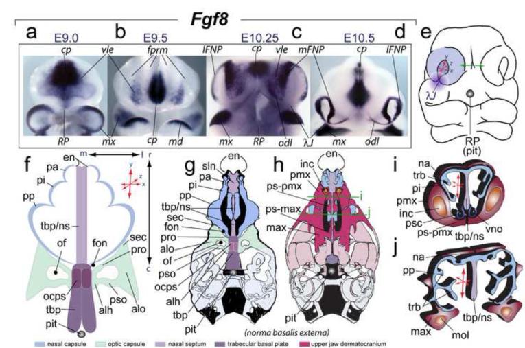
FIGURE 1.
Elaboration of frontonasal Fgf8 expression, its relation to the λ-junction, and the structural orientation and polarity of the frontonasal skeleton. (a-d) In situ hybridization of Fgf8 in the SCE and frontonasal region from E9 to E10.5. The mandibular first arches in ‘a’ and ‘c’ have been removed to better view the ventrolateral ectoderm (vle). (e) Diagram of an E10.5 murine embryo showing inherent polarity (here defined as relative elaboration along the rostro-caudal, medio-lateral and dorso-ventral axes (represented by ‘x:y:z’ coordinate red arrows) of the olfactory pit. Green arrows highlight the fact that contra-lateral mFNPs must eventually conjoin across the midline. Purple gradient disc: central rami of the λ-junction (λJ) (after Compagnucci et al., 2011). Greyscale circle: the position of Rathke’s Pouch (RP) that yields the pituitary (pit) which demarcates the position of caudal boundary of the trabecular basal plate (TBP). (f-i’) Diagrams of the developing skeleton associated with the nasal capsule (NSC), optic capsule (OPC), and midline skeleton of the neurocranium and their orientation and polarity. (f) The three pars of the NSC are in blue, the TBP (midline) structures in shades of purple, and the OPC in green. (g,h) Schemae of norma basalis externa views of neonatal murine skulls. Blue: NSC structure. Purple: TBP midline structures and the Red: upper jaw dermatocranium. Green lines: the relative positions of the coronal sections for “ i ” and “ j ”. (i, j) Nature of NSC polarity as depicted in diagrams of coronal sections of a murine skull.

