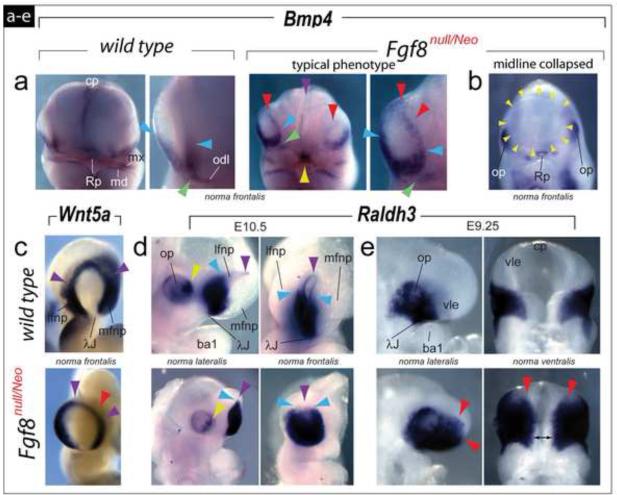
FIGURE 6.
Transformed topography and polarity of regional signaling systems in the frontonasal region of Fgf8null/Neo embryos. (a-e) Comparative in situ hybridization. (a) Although Bmp4 expression at E10.25 is normally restricted to the ventral margins of the OFP at the center of the λ-junction (compare blue arrowheads) and the odontogenic line (odl), in typical Fgf8null/Neo embryos Bmp4 expression extends along the entire rim of the mutant OFP (red arrowheads). Mutant embryos evince a break in Bmp4 expression between the odl and the center of the λ-junction (compare green arrowheads). Purple arrowhead: decreased expression in the mutant commissural plate (cp). Yellow arrowhead: expression in Rathke’s pouch. (b) Lack of detectable Bmp4 transcripts in the OFP in embryos exhibiting a single, collapsed OFP (yellow arrowheads). (c) Wnt5a transcripts are still detected along the rim of the OFP (purple arrowheads) of mutant embryos, though they are diminished in the FNP core (red arrowhead). (d) At E10.5 expression of Raldh3 is diminished in the optic primordia (compare yellow arrowheads) and expanded dorsally within the OFP of mutant embryos (compare the purple arrowheads, indicating the dorsal rim of the pits, and the blue arrowheads, highlighting the dorsal-most extent of extensive Raldh3). (e) At E9.25, Raldh3 is expanded rostrally (red arrowheads) and medially (double headed black arrow) in mutant embryos.

