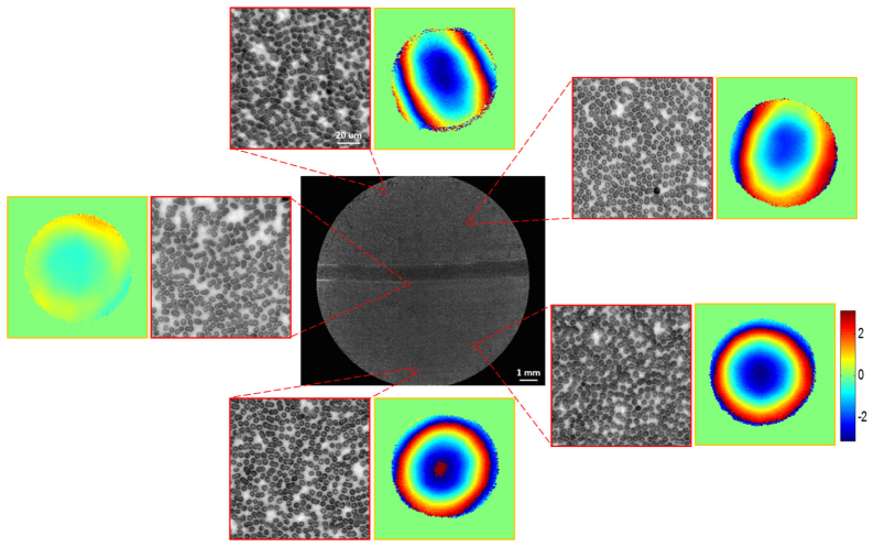
Fig. 4.
Full FOV high resolution monochrome image (red LED illumination) reconstruction of blood smear: the entire FOV is segmented into small tiles, and the aberration is treated as constant in each tile. EPRY-FPM algorithm is run on each tile and the reconstructed high resolution images are mosaicked together. The insets show the detail of the reconstructed image and also the wavefront aberration at those locations.

