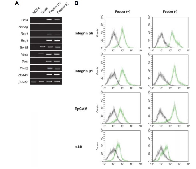
Fig. 2.

Cellular and molecular characterization of SSCs cultured on Matrigel. (A) RT-PCR analysis of SSCs-specific gene expression. Lane 1, MEFs; lane 2, testis; lane 3, SSCs cultured on mitomycin C-treated MEFs, lane 4; SSCs cultured on Matrigel. (B) FACS analysis of the expression of SSC surface proteins: integrin α6 (CD49f), integrin β1 (CD29), EpCAM (CD326), and c-kit (CD117). Black lines, isotype controls; green lines, SSC surface proteins. SSCs are positive for the SSC markers integrin α6 (CD49f), integrin β1 (CD29), EpCAM (CD326) and c-kit (CD117).
