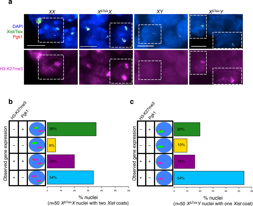
Figure 6. Disassociation of Xist induction, H3-K27me3 enrichment, and inactivation of the XΔTsix maternal X-chromosome in E6.5 extra-embryonic cells.
(a) RNA FISH detection of Xist, Tsix, and Pgk1 RNAs coupled with immunofluorescence (IF) detection of H3-K27me3 in extra-embryonic cells of E6.5 embryos. Dashed boxes mark representative nuclei. Scale bar, 10 µm. (b) Quantification of H3-K27me3 enrichment and Pgk1 expression in nuclei displaying Xist RNA coating of both X-chromosomes in XΔTsixX extra-embryonic cells (50 nuclei with Xist RNA coating of both X-chromosomes were analyzed [n=5 XΔTsixX embryos]). Wild-type (WT) XX embryos show Xist RNA coating and enrichment of H3-K27me3 on a single X-chromosome (n=5 embryos). (c) Quantification of H3-K27me3 enrichment and Pgk1 expression in nuclei displaying Xist RNA coating of the X-chromosome in XΔTsixY extra-embryonic cells (50 nuclei with Xist RNA coating of the single X-chromosome [n = 4 XΔTsixY embryos] were analyzed). WT XY cells show neither Xist RNA coating nor H3-K27me3 enrichment (n = 4 embryos).

