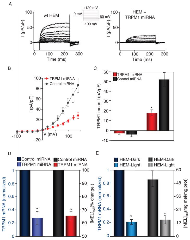
Fig. 4.
Reduced abundance of TRPM1 mRNA correlates with decreased TRPM1 current amplitude and reduced pigmentation in primary HEM cells. (A) Representative endogenous whole-cell currents recorded from wild-type (wt, left) or TRPM1-targeting miRNA-transfected HEMs in response to a voltage step protocol (upper middle). (B) I-V relations of HEMs transfected with scrambled miRNA (control, black circles, n = 6) or anti-TRPM1 miRNA (TRPM1 miRNA, red circles, n = 6). (C) Mean inward (−100 mV) and outward (+140 mV) current recorded in response to a step protocol from HEMs expressing control (scrambled) miRNA and TRPM1 miRNA. The TRPM1 outward current was ~66% smaller in the TRPM1 miRNA–expressing cells (17.9 ± 3.1 pA/pF, n = 6, compared to 52.0 ± 7.1 pA/pF, n = 6 for control miRNA–expressing cells). *P < 0.05; t test. Error bars, ± SEM. (D) HEM-dark cells treated with TRPM1 miRNA (purple) have decreased TRPM1 mRNA abundance when compared to those treated with scrambled miRNA (dark blue) (n = 4 independent experiments). Decreased TRPM1 mRNA abundance correlates with decreased cellular melanin concentration (right axis, n = 4 independent experiments). *P < 0.05; t test. Error bars, ± SEM. (E) In HEM-dark and HEM-light cells, the endogenous abundance of TRPM1 mRNA (left axis) closely correlates with the melanin concentration (right axis, n = 3 independent experiments). An ~80% lower abundance of TRPM1 mRNA in HEM-light compared to HEM-dark cells corresponds to an ~75% reduction in the cellular melanin concentration (n = 3 independent experiments). *P < 0.05; t test. Error bars, ± SEM.

