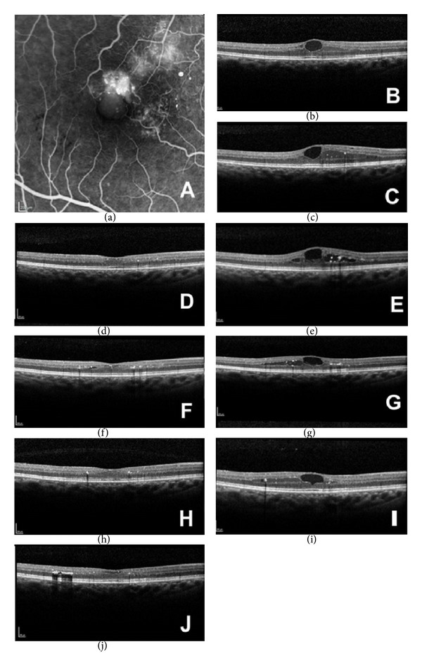
Figure 1.

(a) Fundus fluorescein angiography showing perifoveal macular telangiectasia. (b) Ocular coherence tomography (OCT) of the right eye at presentation showing cystoid macular oedema, CMT = 353 μm. (c) OCT of the right eye on clinic visit following a course of three intravitreal bevacizumab injections (one each consecutive month) showing cystoid macular oedema, CMT = 401 μm. (d) OCT of the right eye on clinic visit following intravitreal triamcinolone injection showing no cystoid macular oedema, CMT = 282 μm. (e) OCT of the right eye on second clinic visit following intravitreal triamcinolone injection showing recurrence of cystoid macular oedema, CMT = 397 μm. (f) OCT of the right eye on clinic visit following intravitreal injection of Ozurdex implant showing reduced cystoid macular oedema, CMT = 286 μm. (g) OCT of the right eye on second clinic visit following intravitreal injection of Ozurdex implant showing a recurrence of cystoid macular oedema, CMT = 349 μm. (h) OCT of the right eye on clinic visit following second intravitreal injection of Ozurdex implant showing a reduction in cystoid macular oedema, CMT = 279 μm. (i) OCT of the right eye on clinic visit at four and a half months following second intravitreal injection of Ozurdex implant showing a recurrence of cystoid macular oedema, CMT = 332 μm. (j) OCT of the right eye on clinic visit at two weeks following a third intravitreal injection of Ozurdex implant showing reduced cystoid macular oedema, CMT = 279 μm.
