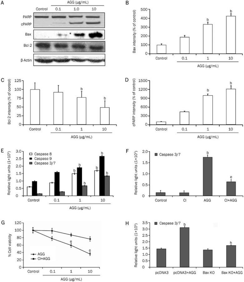
Figure 3.
AGG induces a change in the apoptotic proteins in HepG2 cells. (A) HepG2 cells were treated with the indicated doses of AGG for 24 h, and the cell lysates were immunoblotted with antibodies, as mentioned in 'Methods'. The protein band intensities of Bax (B), Bcl-2 (C) and cleaved PARP (cPARP) (D) were quantified using the ImageJ software with the obtained Western blots of the HepG2 cells. (E) The caspase-Glo 3/7, 8 and 9 activity assays were performed 24 h after AGG treatment with the indicated doses. (F) The HepG2 cells were pre-treated with 50 μmol/L caspase 3 inhibitor (CI) for 2 h prior to AGG (10 μg/mL) exposure for a period of 24 h, and the caspase 3/7 activity was measured as previously mentioned. (G) The viability of the HepG2 cells was measured using a MTT assay with or without the prior addition of CI (as mentioned above) in the AGG (10 μg/mL)-exposed cells for 72 h. (H) The caspase 3/7 activity was also measured in a similar manner in the AGG-treated or untreated HepG2 cells that were transfected with pcDNA3 (mock) or a Bax KO vector, as mentioned in 'Methods'. All data are represented as the mean±SD of three different observations (using Student's t-test). bP<0.05 vs control, eP<0.05 vs AGG treated group and hP<0.05 vs mock transfected AGG treated group, were considered significant.

