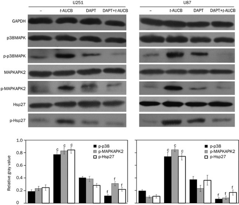
Figure 4.
DAPT blocks t-AUCB-induced activation of the p38 MAPK/MAPKAPK2/Hsp27 pathway. The activation of p38 MAPK, MAPKAPK2, and Hsp27 was detected by Western blotting using antibodies against phosphorylated p38 MAPK (Thr180/Tyr182), phosphorylated MAPKAPK2 (Thr334), and phosphorylated Hsp27 (Ser78). The total protein levels for each were also measured. GAPDH served as a loading control. The relative gray values of p-p38/p38, p-MAPKAPK2/MAPKAPK2, and p-Hsp27/Hsp27 were calculated with the ImageJ program using their measured gray values and represented the phosphorylation levels of p38 MAPK, MAPKAPK2, and Hsp27. In the vehicle control (−), the phosphorylation levels of p38 MAPK, MAPKAPK2, and Hsp27 were very low. After the 48-h treatment with 200 μmol/L t-AUCB, the phosphorylation levels of p38 MAPK, MAPKAPK2, and Hsp27 increased significantly. In cells pre-treated with DAPT before the treatment of t-AUCB, the phosphorylation levels were very low, with no significant change compared to the vehicle control, thus indicating that DAPT blocks the t-AUCB-induced activation of the p38 MAPK/MAPKAPK2/Hsp27 pathway. cP<0.01 vs vehicle control. fP<0.01 vs t-AUCB.

